


The Soft Tissue Oracle: Magnetic Resonance Imaging in Orthopaedics and Sports Medicine
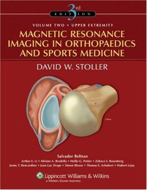
While X-rays are excellent for bone, they are virtually blind to the critical soft tissues that actually stabilize the joints: ligaments, tendons, meniscus, and labrum. Magnetic Resonance Imaging (MRI) revolutionized orthopedics by illuminating this hidden anatomy. Magnetic Resonance Imaging in Orthopaedics and Sports Medicine (2 Volume Set) – 3rd Edition is the massive, universally revered masterwork that defines the interpretation of musculoskeletal MRI. This volume provides the exact, highly advanced blueprints required to read scans with the precision of a subspecialist.
Mastering the Complex Anatomy
The core philosophy of this text is profound anatomical correlation. The authors aggressively pair high-resolution MRI slices with actual cadaveric cross-sections. It dictates exactly how to track the complex, twisting fibers of the Anterior Cruciate Ligament (ACL) across sagittal, coronal, and axial planes, ensuring the reader can confidently differentiate a partial tear from a mucoid degeneration.
Navigating the Postoperative Joint
The book provides a masterclass in advanced interpretation. It dictates the exact imaging pathways for evaluating a joint that has already been operated on. It rigorously covers the artifacts created by metallic suture anchors and the altered signal intensity of a healing meniscus repair, preventing the catastrophic error of diagnosing a “re-tear” when the scan is simply showing normal postoperative scar tissue.
Frequently Asked Questions (FAQs)
Is this a basic introductory book for medical students?
Absolutely not. This is a massive, two-volume, highly technical library designed for advanced readers.
Who is the primary audience?
It is the absolute, mandatory definitive reference for Musculoskeletal Radiologists, Orthopedic Sports Medicine Surgeons, and Radiology Residents.