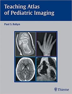

A Visual Masterclass: Teaching Atlas of Pediatric Imaging
Radiology cannot be learned by reading text; it must be learned by analyzing thousands of images until pattern recognition becomes instantaneous. Teaching Atlas of Pediatric Imaging is the definitive, high-yield, fiercely visual casebook designed to force-feed radiological expertise into the reader’s brain. For radiology residents preparing for board exams and pediatricians demanding a deeper understanding of imaging, this volume provides the absolute, uncompromising blueprints required to conquer pediatric pathology.
Case-Based Radiological Learning
The core philosophy of this text is aggressive pattern recognition. The authors discard long, boring chapters and instead present the reader with unknown, high-quality images—just like a real clinical shift or a terrifying oral board exam. The reader is forced to look at the MRI, CT, or X-ray, commit to a diagnosis, and then turn the page to face the brutal reality of whether they were right or dangerously wrong.
Deconstructing Complex Pediatric Pathology
The book provides a masterclass in pathological differentiation. Once the reader flips the page, the authors ruthlessly deconstruct the image. They provide the exact “Aunt Minnie” (classic, unmistakable) radiological signs of specific diseases. They detail exactly why that specific shadow on the pediatric chest X-ray is a neuroblastoma and not a benign cyst, providing the rigid diagnostic criteria required to definitively rule out lethal conditions.
Musculoskeletal Trauma and Skeletal Dysplasias
This is a fiercely practical guide to the pediatric skeleton. The text provides massive sections on pediatric bone imaging. It dictates exactly how to identify subtle Salter-Harris growth plate fractures that, if missed, will result in permanent limb deformity. It also covers the terrifyingly complex world of skeletal dysplasias (genetic bone disorders), providing the exact radiological measurements required for diagnosis.
High-Yield Board Review and Unknown Cases
The final value of this reference is its absolute focus on passing exams and surviving the reading room. The cases are explicitly chosen because they are highly tested on radiology board exams and frequently missed in clinical practice. The authors provide the exact “pearls and pitfalls” for each case, ensuring the resident never falls for a classic radiological trick.
Frequently Asked Questions (FAQs)
Is this a comprehensive textbook?
No, it is an Atlas. It is designed for rapid, visual, case-based learning, not for reading exhaustive chapters on the cellular biology of diseases.
Does it cover all imaging modalities?
Yes, the cases span plain film radiography (X-ray), Fluoroscopy, Ultrasound, Computed Tomography (CT), and Magnetic Resonance Imaging (MRI).
Who is the primary audience?
It is the absolute, undisputed, mandatory study guide for Radiology Residents, Pediatric Radiology Fellows, and attending Pediatricians.
Conclusion
Teaching Atlas of Pediatric Imaging proves that clinical brilliance in radiology is born from relentless visual practice. It is an indispensable survival guide for board exams and overnight hospital shifts. By grinding through these complex unknown cases, mastering the “Aunt Minnie” signs, and absorbing the rigid diagnostic pearls detailed in this volume, physicians build the ironclad pattern recognition required to never miss a pediatric diagnosis.