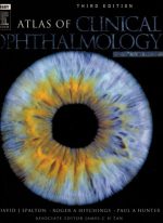


The Visual Encyclopedia: Atlas of Clinical Ophthalmology
In ophthalmology, a diagnosis is rarely made by a blood test; it is made by pattern recognition. The clinician must look through a microscope and instantly identify the pathology based solely on its visual appearance. Atlas of Clinical Ophthalmology is a fiercely comprehensive, heavily illustrated masterwork designed to train the clinician’s eye across every subspecialty. This volume provides the exact visual blueprints required to recognize blinding diseases before they progress.
Mastering the Anterior Segment
The core philosophy of this text is high-resolution correlation. The authors aggressively pair pristine slit-lamp photography with concise clinical descriptions. It dictates exactly how to recognize the classic “dendritic” (branching) ulcer of a Herpes Simplex virus infection on the cornea, a critical pattern that must be identified instantly, as treating it with standard steroid drops will cause the virus to multiply and melt the eye.
Navigating the Fundus
The book provides a masterclass in posterior pole pathology. It dictates the exact visual pathways for identifying retinal disease. It rigorously displays the evolution of diabetic retinopathy, training the reader to spot the microscopic red dots (microaneurysms) of early disease, and the terrifying, frond-like webs of bleeding new blood vessels (neovascularization) that indicate the eye is actively going blind.
Frequently Asked Questions (FAQs)
Does this book provide detailed surgical instructions?
No, it is a *diagnostic atlas* focused entirely on the visual presentation and clinical identification of eye diseases.
Who is the primary audience?
It is the absolute, mandatory visual reference for Ophthalmology Residents, Optometry Students, and Comprehensive Ophthalmologists.