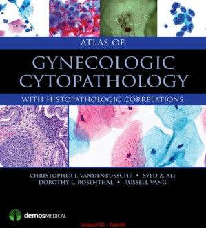



The Microscopic Verdict: Atlas of Gynecologic Cytopathology
A surgeon cannot remove a woman’s uterus based on a suspicion; they must have absolute, undeniable microscopic proof that cancer exists. The pathologist is the final judge. Atlas of Gynecologic Cytopathology: With Histopathologic Correlations is the brutally precise, fiercely visual master text detailing exactly how to decode the cellular architecture of the female reproductive tract. This volume provides the exact histological blueprints required to translate a smear of cells on a glass slide into a definitive, life-altering diagnosis.
Mastering the Cellular Aberration
The core philosophy of this text is pattern recognition at the microscopic level. The authors aggressively detail the mechanics of the Pap test. It dictates exactly how a pathologist must identify the darkened, enlarged nucleus of a “koilocyte,” mathematically proving the presence of Human Papillomavirus (HPV) and instantly categorizing the patient’s risk for developing invasive cervical cancer.
Navigating the Histopathologic Correlation
The book provides a masterclass in verifying the diagnosis. It dictates the exact visual pathways for confirming cytology with actual tissue biopsies. It rigorously explores how to match the free-floating cells of a fine-needle aspiration with the solid architectural structure of an actual tissue sample, ensuring that the diagnosis of a high-grade sarcoma is absolutely correct before the patient is subjected to massive, radical surgery.
Frequently Asked Questions (FAQs)
Does this book teach doctors how to prescribe chemotherapy?
No, it is a strictly specialized *anatomic pathology, cytology, and laboratory diagnostic atlas* focused entirely on looking through a microscope.
Who is the primary audience?
It is the absolute, mandatory visual guide for Cytopathologists, Surgical Pathologists, Cytotechnologists, and Pathology Residents.