
The Visual Library: Color Atlas and Synopsis – Wills Eye – Retina
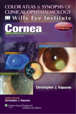
The retina is the neurosensory wallpaper of the eye; diagnosing its diseases requires absolute pattern recognition. A clinician cannot treat a retinal detachment or a macular hole if they do not know exactly what it looks like through an indirect ophthalmoscope. Color Atlas and Synopsis of Clinical Ophthalmology – Wills Eye Institute – Retina is a fiercely visual, heavily illustrated masterwork designed to rapidly train the clinician’s eye. This volume provides the exact, high-resolution photographic blueprints required to instantly recognize posterior segment pathology.
Mastering Retinal Morphology
The core philosophy of this text is that seeing is diagnosing. The authors aggressively pair massive, full-color fundus photographs with concise clinical summaries. It dictates exactly how to differentiate the “cherry-red spot” of a Central Retinal Artery Occlusion (a devastating stroke of the eye) from the “blood and thunder” hemorrhages of a Central Retinal Vein Occlusion.
Navigating Advanced Imaging
The book provides a masterclass in diagnostic correlation. It dictates the exact clinical pathways for utilizing Optical Coherence Tomography (OCT) and Fluorescein Angiography (FA). It rigorously pairs the clinical photograph of a diabetic macular edema with its corresponding OCT cross-section, showing exactly how the intraretinal fluid physically separates the photoreceptor layers, causing blindness.
Frequently Asked Questions (FAQs)
Is this a massive, text-heavy encyclopedic reference?
No, it is an “Atlas and Synopsis”—relying heavily on high-quality *images* paired with highly condensed, bullet-pointed clinical facts for rapid pattern recognition.
Who is the primary audience?
It is the absolute, mandatory visual guide for Ophthalmology Residents, Optometrists, and General Ophthalmologists managing retinal disease.