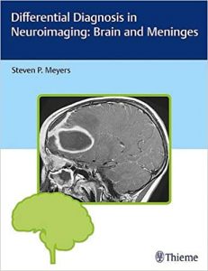


Authored by renowned neuro-radiologist Steven P. Meyers, Differential Diagnosis in Neuroimaging: Brain and Meninges is a stellar guide for identifying and diagnosing brain pathologies based on location and neuroimaging results. The succinct text reflects more than 25 years of hands-on experience gleaned from advanced training and educating residents and fellows in radiology, neurosurgery, and neurology. The high-quality MRI, CT, PET, PET/CT, conventional angiography, and X-ray images have been collected over Dr. Meyers’s lengthy career, presenting an unsurpassed visual learning tool.
The distinctive ‘three-column table plus images’ format is easy to incorporate into clinical practice, setting this book apart from larger, disease-oriented radiologic tomes. The layout enables readers to quickly recognize and compare abnormalities based on high-resolution images.
Key Highlights
- Tabular columns organized by anatomical abnormality include brain imaging findings and a summary of key clinical data that correlates to the images
- Comprehensive imaging of the brain, ventricles, meninges, and neurovascular system in both children and adults, including congenital/developmental anomalies and acquired disease
- More than 1,900 figures illustrate the radiological appearance of intracranial lesions, masses, neurodegenerative disorders, ischemia and infarction, and more
This visually rich resource is a must-have diagnostic tool for radiologists, neurosurgeons, and neurologists, and residents and fellows.