
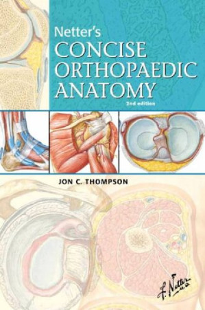

The Visual Library: Netter’s Concise Orthopaedic Anatomy (2nd Edition)
Orthopedic surgery is, at its core, applied, three-dimensional anatomy. A surgeon cannot safely navigate the human body without an absolute, perfect mental map of every muscle, nerve, and artery. Frank Netter’s illustrations are the legendary, universally recognized gold standard of medical art. Netter’s Concise Orthopaedic Anatomy (2nd Edition) takes these iconic paintings and aggressively weaponizes them for the surgical resident. This volume provides the exact, visually stunning blueprints required to memorize the surgical anatomy of the human body.
Mastering the Visual Space
The core philosophy of this text is that visual memory is superior to text. The authors aggressively pair Netter’s paintings with high-yield clinical tables. It dictates exactly how to visualize the brachial plexus, turning a confusing jumble of nerves into a perfectly clear, color-coded diagram of roots, trunks, divisions, cords, and branches, making it impossible to forget.
Navigating Surgical Correlation
The book provides a masterclass in anatomical relevance. It dictates the exact clinical pathways where anatomy dictates pathology. It rigorously details the precise blood supply to the talus, visually explaining why a fracture through the neck of the bone almost always results in avascular necrosis (AVN) of the body, as the retrograde blood vessels are sheared off by the injury.
Frequently Asked Questions (FAQs)
Is this a massive textbook full of long, dense chapters?
No, it is a “Concise” atlas. It relies heavily on illustrations, tables, and bullet points for rapid memorization and review.
Who is the primary audience?
It is the absolute, mandatory anatomical atlas for Orthopedic Residents (especially interns), Medical Students on surgical rotations, and Physician Assistants.


