
The Radiographic Translator: Orthopedic Imaging: A Practical Approach
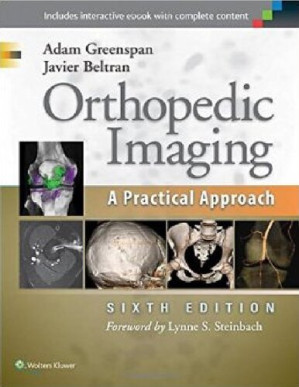
Before a surgeon can reconstruct a joint or fix a shattered bone, they must first perfectly understand the three-dimensional geometry of the injury hidden beneath the skin. Orthopedic Imaging: A Practical Approach (6th Edition) is a fiercely pragmatic, highly comprehensive manual designed to seamlessly bridge the gap between clinical orthopedics and diagnostic radiology. This volume provides the exact blueprints required to independently interpret complex musculoskeletal scans.
Mastering Modality Integration
The core philosophy of this text is knowing exactly what to order and why. The authors aggressively detail the strengths and weaknesses of different imaging technologies. It dictates exactly why a CT scan is mandatory for surgical planning in a complex tibial plateau fracture (to map the exact location of the depressed articular fragments), while an MRI is required to evaluate the associated meniscal and ligamentous tears.
Navigating Subtle Pathology
The book provides a masterclass in detecting the easily missed lesion. It dictates the exact anatomical pathways for reading trauma films. It rigorously details how to identify the “segond fracture” (a tiny fleck of bone off the lateral tibia) on a standard AP knee X-ray, which is pathognomonic for a massive, catastrophic rupture of the Anterior Cruciate Ligament (ACL).
Frequently Asked Questions (FAQs)
Is this a book purely for radiologists?
No, it is specifically designed to be “Practical” for the *ordering clinician*—the orthopedic surgeon or sports physician who needs to read the scan before the official report is dictated.
Who is the primary audience?
It is the absolute, mandatory imaging guide for Orthopedic Residents, Sports Medicine Physicians, and Musculoskeletal Radiologists.