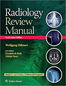

The Definitive Imaging Encyclopedia: Radiology Review Manual (Sae) (Pb 2021)
In the high-stakes, terrifyingly fast-paced world of diagnostic radiology, missing a subtle 3-millimeter hyperdensity on a CT scan isn’t a minor error—it’s the difference between catching a curable tumor and missing a fatal cancer. Radiology Review Manual is the definitive, fiercely optimized, and globally renowned “survival bible” for radiology residents. For doctors preparing for their grueling board exams, this massive volume provides the absolute, uncompromising data compression required to instantly recognize thousands of different pathologies across every single imaging modality.
The Ultimate High-Yield Radiology Board Review
The core philosophy of this text is radical data compression and extreme speed. The authors understand that a radiology resident must memorize an impossible amount of information. The book completely strips away long, flowing paragraphs of text. It is structured entirely as an aggressive, high-yield outline. It utilizes massive bulleted lists, rapid-fire differential diagnosis tables, and thousands of specific “radiologic signs” designed for instant visual recall.
Deconstructing Musculoskeletal and Neurological Pathology
The book provides a masterclass in full-body diagnostics. The authors aggressively categorize the human body by system. Readers will find exhaustive outlines detailing exactly how to instantly differentiate a benign enchondroma from a lethal chondrosarcoma on a bone X-ray. The neuroradiology sections provide the exact, rigid MRI signal characteristics (T1, T2, FLAIR) required to instantly diagnose a terrifying glioblastoma multiforme or a subtle multiple sclerosis plaque.
Mastering Complex MRI, CT, and Advanced Ultrasound
This is a fiercely multi-modal publication. The book does not just teach anatomy; it teaches the physics of the image. The authors provide the exact protocols for analyzing everything from a massive, multi-phase contrast CT of the liver (to detect hepatocellular carcinoma) to the highly nuanced grayscale subtleties of a thyroid ultrasound, ensuring the resident can navigate any imaging study ordered by the emergency room.
Rapid Differential Diagnosis Based on Subtle Imaging Signs
The final value of this reference is its legendary focus on “Gamuts” (differential diagnoses). If a resident sees a “ground-glass opacity” in the lungs, the book provides the exact, prioritized list of every single disease that can cause it, and provides the exact secondary clues required to eliminate the wrong answers and arrive at the singular, correct diagnosis.
Frequently Asked Questions (FAQs)
Does this book contain thousands of image examples?
Actually, no. This is the unique genius of the “Radiology Review Manual.” It is primarily a text-based outline designed to be used alongside your PACS system or image atlases. It organizes the data in your brain so you know what you are looking for when you see the actual image.
Is this useful for non-radiologists?
It is highly useful for specific specialists (like orthopedic surgeons or neurologists) who need a rapid, high-yield reference for interpreting the imaging of their specific organ systems.
Who is the primary audience?
It is the absolute, mandatory, undisputed “bible” for Radiology Residents preparing for their CORE and Certifying Board Exams worldwide.
Conclusion
Radiology Review Manual proves that surviving radiology board exams requires absolute, militaristic organization of clinical data. It is an indispensable tool for the dark reading room. By utilizing the aggressive outlines, the rapid-fire differential tables, and the massive data compression provided in this volume, residents can process imaging studies with flawless speed and accuracy, guaranteeing their success as attending diagnostic radiologists.