

Mastering Acoustic Impedance: Textbook of Diagnostic Sonography (Volume 2)
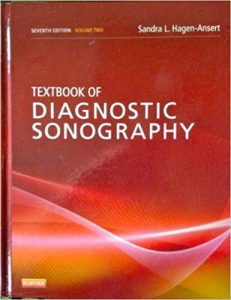
An ultrasound machine is not a camera; it is an incredibly complex radar system. It sends high-frequency sound waves into the human body, and the sonographer must instantly interpret the echoes bouncing back off moving organs, flowing blood, and fetal structures. Textbook of Diagnostic Sonography (Volume 2) is the definitive, fiercely advanced clinical manual dedicated entirely to the mastery of this highly operator-dependent modality. For diagnostic medical sonographers, radiologists, and obstetricians, this massive volume provides the uncompromising blueprints required to extract flawless diagnostic data using nothing but sound.
The Definitive Guide to Advanced Ultrasound Imaging
The core philosophy of this text is absolute technical and anatomical precision. The authors understand that an ultrasound is only as good as the person holding the probe. While Volume 1 covers the basics and the abdomen, Volume 2 aggressively focuses on the most complex, high-stakes areas of sonography: Obstetrics, Gynecology, and advanced Vascular hemodynamics.
Deep Dive into Obstetric and Gynecologic Sonography
The book provides a masterclass in fetal diagnostics. The authors dedicate massive sections to the terrifying responsibility of obstetric ultrasound. They provide the exact, rigid protocols for measuring fetal biometry to determine gestational age. More importantly, they detail the highly advanced, slice-by-slice anatomical surveys required to detect catastrophic fetal anomalies—from severe congenital heart defects to terrifying neural tube defects (spina bifida)—long before the child is born.
Vascular Ultrasound, Doppler Mechanics, and Hemodynamics
This is a fiercely physics-based diagnostic guide. The authors aggressively tackle the complexities of vascular imaging. They break down the physics of the Doppler shift. Readers will learn exactly how to interpret the color flow and spectral waveforms of the carotid arteries and the deep veins of the legs, providing the exact mathematical velocities required to instantly diagnose a massive deep vein thrombosis (DVT) or a stroke-causing arterial stenosis.
The Physics of Artifacts and Image Optimization
The final value of this reference is its focus on physics and troubleshooting. The authors detail the exact buttons on the machine required to fix a terrible image. They teach the sonographer how to manipulate gain, focal zones, and frequency to penetrate deep tissue, and how to instantly recognize “artifacts” (like acoustic shadowing from a gallstone or reverberation from air) that can mimic or obscure real disease.
Frequently Asked Questions (FAQs)
Do I need Volume 1 to understand Volume 2?
Volume 1 covers the foundational physics and general abdominal imaging. Volume 2 assumes that basic knowledge and focuses entirely on advanced Obstetrics, Gynecology, and Vascular imaging. Both are typically required for full board certification.
Does it cover echocardiography (heart ultrasound)?
While it covers fetal echocardiography and general vascular hemodynamics, dedicated adult cardiac echocardiography is usually covered in specialized cardiology texts.
Who is the primary audience?
It is the absolute, mandatory curriculum for Diagnostic Medical Sonography students preparing for their ARDMS registries, as well as Maternal-Fetal Medicine (MFM) specialists and Radiologists.
Conclusion
Textbook of Diagnostic Sonography proves that finding disease with sound requires absolute mastery of both physics and anatomy. It is an indispensable tool for the ultrasound suite. By mastering the complex Doppler hemodynamics, the rigorous obstetric scanning protocols, and the image optimization physics detailed in this volume, sonographers become the highly skilled “eyes” of the diagnostic team, capable of catching life-threatening diseases in real-time.