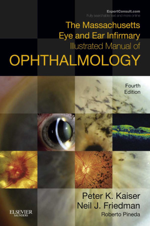



The Visual Triage: Massachusetts Eye and Ear Infirmary Illustrated Manual (4th Ed)
When triaging a patient, text descriptions of ocular pathology often fail to capture the subtle nuances that differentiate a benign condition from a blinding emergency. A picture is not just worth a thousand words; it is often the entire diagnosis. The Massachusetts Eye and Ear Infirmary Illustrated Manual of Ophthalmology (4th Edition) is a fiercely visual, heavily illustrated clinical atlas designed specifically for rapid diagnostic correlation. This volume provides the exact visual blueprints required to match the patient’s eye to the textbook pathology.
Mastering the Anterior Chamber
The core philosophy of this text is immediate pattern recognition. The authors aggressively pair high-resolution slit-lamp photography with the corresponding diagnosis. It dictates exactly how to look at a photograph of a hyphema (blood layering in the front of the eye) following trauma, and instantly recognize the “eight-ball” appearance that indicates the drain is completely clogged and the eye pressure is about to skyrocket to catastrophic levels.
Navigating the Fundus
The book provides a masterclass in posterior segment imaging. It dictates the exact visual pathways for identifying retinal emergencies. It rigorously trains the clinician to recognize the pale, swollen retina and the classic “cherry-red spot” of a Central Retinal Artery Occlusion, ensuring the patient is immediately rushed for a stroke workup rather than being sent home with a misdiagnosis.
Frequently Asked Questions (FAQs)
How does this differ from the MEEI “Review Manual”?
While the Review Manual is text-heavy and focused on board preparation, this *Illustrated Manual* is heavily visual, acting as a direct atlas and clinical comparison tool.
Who is the primary audience?
It is the absolute, mandatory visual guide for Emergency Room Physicians, Optometrists, Medical Students, and first-year Ophthalmology Residents.